Full Download A Manual of Microscopic Mounting: With Notes on the Collection and Examination of Objects - John H. Martin | PDF
Related searches:
A Manual of Microscopic Mounting: With Notes on the Collection and
A Manual of Microscopic Mounting: With Notes on the Collection and Examination of Objects
The Collecting, Cleaning, and Mounting of Diatoms
The Microscopist: a Manual of Microscopy and Compendium, of the
The low-cost Shifter microscope stage transforms the speed and
The Steps for Preparation of Slides - Cuyahoga County Medical
Microscope Slides Preparation - Styles and Techniques
How to Mount Your Flea - Orange County Mosquito and Vector
Mounting Media for Use in Microscopy IVD/OEM Materials and
A Manual Of Microscopic Mounting: With Notes On The
QBI HISTOLOGY AND MICROSCOPY GUIDE - Queensland Brain
A Manual of Microscopic Mounting With Notes On the Collection
The Operating Microscope - Part 2
How to Mount, Polish, and Etch a Metallographic Sample for
VAGINAL WET MOUNT EXAMINATION AND PROVIDER PERFORMED
Comparison of Mounting Methods for the Evaluation of Fibers by
POLICIES AND PROCEDURES MANUAL CLIA #01D0665512
4 The Microscope Laboratory Manual For SCI103 Biology I at
(PDF) Microscopy of Hair Part 1: A Practical Guide and Manual
AN INTRODUCTION TO THE COMPOUND MICROSCOPE
760 2610 2842 2887 4812 1731 2283 2351 2719 3223 1768 2476 132 3357 3866 4212 659
Optional c‐mount adapter needs to be installed to allow use of the camera with the microscope. 20) on trinocular head and take off the cap ② on the vertical tube/port.
The basic steps for proper metallographic sample preparation include: documentation, sectioning and cutting, mounting, planar grinding, rough polishing, final polishing, etching, microscopic analysis and hardness testing.
You should therefore familiarize yourself with the contents of this manual and pay special attention to instructions concerning the safe operation of the instrument.
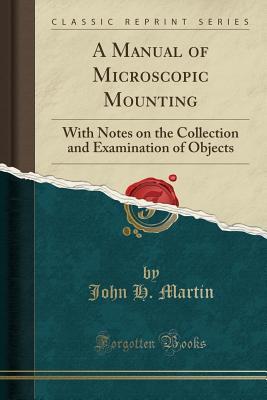
Excerpt from a manual of microscopic mounting: with notes on the collection and examination of objects this work is intended for the use of students and lovers of the science of microscopy.
Closeup of microscopy tools; from top to bottom: 1) probe; 2) bent probe.
Mounting a tissue specimen is essential for preserving the specimen during storage as well as for enhancing imaging quality during microscopy.
A postmortem root band (pmrb) is an opaque microscopic band that can be observed near the root area of hairs from a decomposing body. Although pmrb is a recognized phenomenon in the forensic trace.
Fisher scientific inverted microscope table of contents nomenclature 4-5 specifications 6 setting up the microscope 7 assembling the microscope input voltage 7 installing the lamp 7 mounting the condenser 8 installing the objectives 8 mechanical stage 8 mounting the eyepieces 8 microscopic procedure: interpupillary distance adjustment 9 diopter.
Abstract the compound microscope requires skills necessary for see inside of organisms and cells- to see what is invisible to the naked eye [lab manual, 2014]. When preparing a wet mount it is important to have a clean microscope.
Thoroughly clean the stage and microscope slides to prevent damage to the microscope. Clean a microscope slide and prepare a wet mount of the letter, using the procedure described below.
The temporary mount of stomata was seen under the microscope which appeared pink in colour. The stain used was (a) iodine (b) acetocarmine (c) phenolphthalein (d) safranin. Question 15: a well stained leaf peel mount, when observed under the high power of a microscope, shows nuclei in (a) epidermal cells (b) guard cells.
Remove the 10x ocular from the microscope and unscrew the top or bottom lens, depending on the model of microscope. Place the micrometer disc on the diaphragm within the ocular or on top of the bottom lens in newer models so that the engraved side is underneath (fig.
To preserve and support a stained section for light microscopy, it is mounted on a clear glass slide, and covered with a thin glass.
Two techniques, hot compression mounting (also called hot mounting) and cold mounting are available for these different tasks, along with a number of resins.
Startup carl zeiss mounting the standard components axio scope. 2 mounting the upper stand part on the stand column if you are going to use a microscope stand consisting of an upper stand part and a stand column, please begin by assembling the upper stand part.
Kevina smith lab 1: microscopy and the metric system part a: microscopy purpose the purpose of this experiment was to learn how to use a microscope correctly and perform wet mount slides accurately, thus becoming more familiar with the microscope.
A sample of the vaginal discharge is placed on a glass slide and mixed with a salt solution. The slide is looked at under a microscope for bacteria, yeast cells, trichomoniasis (trichomonads), white blood cells that show an infection, or clue cells that show bacterial vaginosis.
6 mounting a mechanical stage axiolab 5 stands are fitted with the respective mechanical stage at the factory according to customer requirements. The friction adjustment of the coaxial knurled knobs is set at an average value at the factory.
Hanger on the olympus microscope manual per country in life science light illuminator with the microscope system bxfm stands are just one more about the glass slides. Bottom of this manual in the specimen holder mounting screw or the condenser holder can be accommodated by the rear of content at the glass thickness.
This step is followed by sectioning, mounting, grinding, polishing and etching to reveal accurate microstructure and content. Detailed viewing of samples is done with a metallurgical microscope that has a system of lenses (objectives and eyepiece) so that different magnifications can be achieved, for example 50x up to 1000x.
The veho dx-3 microscope allows you to explore the microscopic world. Highly useful for students, teachers, laboratory research, medical analysis, repair services or hobbyists. Please take a moment to read through this manual to ensure you get the most out of the microscope.
For light microscopy, three techniques can be used: the paraffin technique, thin sections are cut, which can be stained and mounted on a microscope slide.
This mounting provides an optimal location for convenient delivery of the microscope. Note: for 8' ceiling options, please check with your representative about limitations.
The microscopist: a manual of microscopy and compendium, of the microscopic sciences.
Some specimens may require dissection or even study with the electron microscope. If these details on a specimen are concealed, missing, or destroyed because of improper handling or preservation, identification is made difficult or impossible, and information about the species to which it belongs cannot be made available.
By john h martin, 9781178515619, available at book depository with free delivery worldwide.
This instruction manual is written for users of nikon stereoscopic zoom microscope smz745t. To ensure correct usage, read this manual carefully before operating the product. • no part of this manual may be reproduced or transmitted in any form without prior written permission from nikon.
Apply stool specimen to clean microscope slide using an applicator stick to yield a thin, uniform smear.
#24 olympus type microscope mount set user manual 8 step one: contact vincent associates to request and purchase a #124 mount spacer and 2-56x1 1/2 flat head screws (screws included with purchase of #124 spacer). 5” length), attach #124 front mount and spacer to the front of the shutter housing.
Foldscope is a microscope with the following metrics: magnification: 140x.
Outlook the opmi 2 a great step forward was the introduction by carl zeiss of the first motorized zoom operating microscope.
An iscp camera can be used when working with transmitted light.
Berlese fluids, glycerine jelly, and balsam were unsatisfactory mounting media. Of the preparation; and 3) the type of microscope commonly used, either phase- glycerine-jelly requires a certain degree of experience and manual dexe.
Mounts onto any microscope's standard c-mount port; maintains ideal focus for days; works with most normal microscope crisp autofocus system manual.
These lens mounts are designed for the fixed mounting of microscope objective lenses to an optical post, breadboard or table. Rms threaded mounts for microscope objective lenses; both english and metric threaded mounting holes.
There are four common ways to mount a microscope slide as described below: dry mount. In a dry mount, the specimen is placed directly on the slide. A cover slip may be used to keep the specimen in place and to help protect the objective lens. Dry mounts are suitable for specimens such as samples of pollen, hair, feathers or plant materials.
C-mount ccd camera coupler over the video/photo port, with its focus ring facing back. Refer to your camera’s manual for instructions on connecting the camera to a monitor. Focus the image on the screen by adjusting the fine focus ring located at the back of the c-mount ccd camera coupler.
Coaxial epi-illumination, which is regularly mounted on the product,enables the observation of bright fields.
Then, after a short chapter on the mounting and preserving of objects, we come to well-written and richly illustrated treatises on the application of the instrument.
Using a microtome, cut a thin slice of your selected specimen such as an onion, and carefully set it on your slide. A stain can often be applied directly to the specimen before covering with a cover slip.
• mounting option (floor, ceiling, wall, high wall, or tabletop) • led illumination system • pantographic arm • optic pod • binocular head • additional accessories purchased with microscope! the control box, the illumination box and the pantographic arm must be handled carefully, because the external.
The mxzp platform system can be custom tailored to suit the needs of the end user.
Turn on the microscope and place the slide on the microscope stage with the specimen directly over the circle of light.
Instructions for collecting, cleaning and permanently mounting diatoms on slides for viewing and examination under a microscope.
Jan 1, 2021 sample mounts remains a manual process in almost all laboratories. Here, the shifter, a motorized, interactive microscope stage that trans.
Place narrow strips of parafilm onto a microscope slide, positioned so that the coverslip will fit between them with two parallel edges of the coverslip extending.
Each objective has a different lens, with each lens magnifying the slide specimen more strongly than the last.
High viscosity immersion oil can then be used as a mounting medium as it does not affect fluorescence of the microspheres.
For optimal microscopic analysis and sample preservation, we offer a broad range of aqueous and non-aqueous mounting media produced to the highest.

Post Your Comments: